
GFP Monoclonal Antibody


ELISA, Flow Cytometry, Immunohistochemistry, IF Microscopy, Western Blot
Mouse
eGFP; rGFP; Wild Type
IgG
Monoclonal
Recombinant Green Fluorescent Protein (GFP) fusion protein corresponding to the full length amino acid sequence (246 aa) derived from the jellyfish Ae
mouse anti-GFP antibody, Green Fluorescent Protein, GFP antibody, Green Fluorescent Protein antibody, EGFP, enhanced Green Fluorescent Protein, Aequorea victoria, Jellyfish


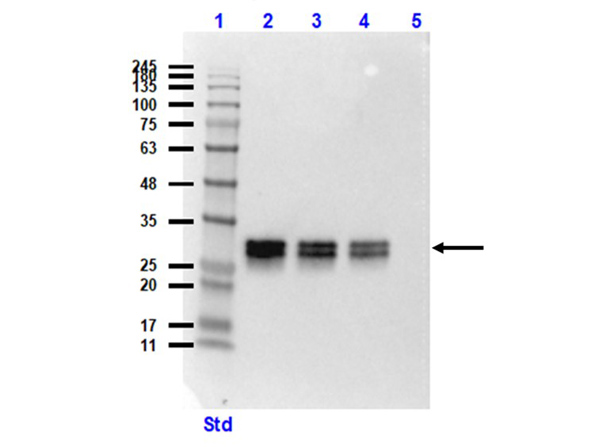
Mouse anti-GFP antibody is functional by western blot, ELISA, Immunofluorescence Microscopy and Immunohistochemistry. Green fluorescent protein is a 27 kDa protein produced from the jellyfish Aequorea victoria, which emits green light (emission peak at a wavelength of 509nm) when excited by blue light. GFP is an important tool in cell biology research. GFP is widely used enabling researchers to visualize and localize GFP-tagged proteins within living cells without the need for chemical staining. Monoclonal anti-GFP is designed to detect enhanced GFP and GFP containing recombinant proteins. Tested in ELISA, IP, and WB and suitable in FACS, IHC, IF. This antibody can be used to detect GFP by ELISA (sandwich or capture) for the direct binding of antigen. Biotin conjugated monoclonal anti-GFP is well suited to titrate GFP in a sandwich ELISA in combination with Rockland's polyclonal anti-GFP (600-101-215) as the capture antibody. Only use the monoclonal form for the detection of enhanced or recombinant GFP. Polyclonal anti-GFP detects all variants of GFP tested to date. The biotin conjugated detection antibody is typically used with streptavidin conjugated HRP (code # S000-03) or other streptavidin conjugates. The use of polyclonal anti-GFP results in significant amplification of signal when fluorochrome conjugated polyclonal anti-GFP is used relative to the fluorescence of GFP alone. For immunoblotting use either alkaline phosphatase or peroxidase conjugated anti-GFP to detect GFP or GFP containing proteins on western blots. Optimal titers for applications should be determined by the researcher.

