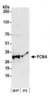
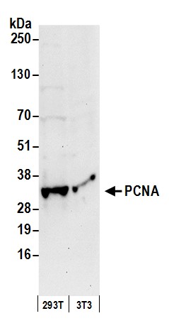
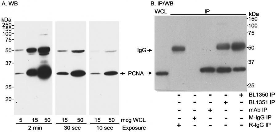
Rabbit anti-PCNA Antibody, Affinity Purified
Antibody was affinity purified using an epitope specific to PCNA immobilized on solid support. The epitope recognized by A300-277A maps to a region between residue 225 and the C-terminus (residue 261) of human proliferating cell nuclear antigen using the numbering given in SwissProt entry P12004 (GeneID 5111). Immunoglobulin concentration was determined by extinction coefficient: absorbance at 280 nm of 1.4 equals 1.0 mg of IgG.
- Data sheet/protocol: View or download
- MSDS: View or download
- Applications: WB, IP, IHC, IHC-IF
Antibodies
ATLD2, cyclin, DNA polymerase delta auxiliary protein, PCNA, proliferating cell nuclear antigen
Human, Mouse
between 225 and C-term
2 - 8°C
Rabbit
Whole IgG
Tris-citrate/phosphate buffer, pH 7 to 8 containing 0.09% Sodium Azide
Polyclonal
12352203

Detection of human and mouse PCNA by western blot. Samples: Whole cell lysate (50 µg) from HEK293T and mouse NIH 3T3 cells prepared using NETN lysis buffer. Antibody: Affinity purified rabbit anti-PCNA antibody A300-277A (lot A300-277A-1) used for WB at 0.05 µg/ml. Detection: Chemiluminescence with an exposure time of 30 seconds.

Detection of human PCNA by western blot and immunoprecipitation. Samples: A. Whole cell lysate (WCL) from A-431 cells. B. WCL (500 µg for IP; 30 µg for WCL lane) from MDA-MB-468 cells. Antibodies: A. Affinity purified rabbit anti-PCNA antibody BL1351 (Cat. No. A300-277A) used at 0.1 µg/ml for WB. B. PCNA was immunoprecipitated using BL1350 (Cat. No. A300-276A), BL1351 or a mouse monoclonal antibody to PCNA (mAb). Control mock IP was performed using normal mouse IgG (M-IgG) and normal rabbit IgG (R-IgG). immunoprecipitatesd PCNA was blotted using a mixture of BL1350 and BL1351 (each at 1 µg/ml). Detection: Chemiluminescence with an exposure times as indicated (A) or 5 seconds (B).

Detection of human PCNA by immunohistochemistry. Sample: FFPE section of human stomach carcinoma. Antibody: Affinity purified rabbit anti- PCNA (Cat. No. A300-277A Lot1) used at a dilution of 1:4,000 (0.25 µg/ml). Detection: Red-fluorescent goat anti-rabbit IgG-heavy and light chain cross-adsorbed Antibody DyLight® 594 Conjugated used at a dilution of 1:100.