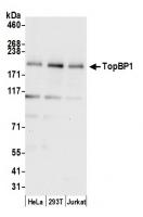
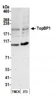
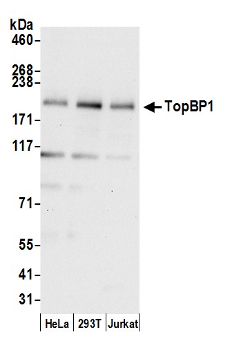
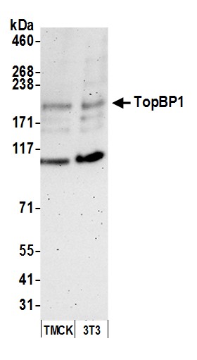
Rabbit anti-TopBP1 Antibody, Affinity Purified - A300-111A - 100 µg - Bethyl Laboratories, Inc. - antibodies


Antibody was affinity purified using an epitope specific to TopBP1 immobilized on solid support. The epitope recognized by A300-111A maps to a region between residue 1400 and the C-terminus (residue 1435) of human Topoisomerase (DNA) II Binding Protein 1 using the numbering given in entry NP_008958.1 (GeneID 11073).Immunoglobulin concentration was determined by extinction coefficient: absorbance at 280 nm of 1.4 equals 1.0 mg of IgG.
Applications:
WB, IP

Detection of human TopBP1 by western blot. Samples: Whole cell lysate (15 µg) from HeLa, HEK293T, and Jurkat cells prepared using NETN lysis buffer. Antibody: Affinity purified rabbit anti-TopBP1 antibody A300-111A (lot A300-111A-2) used for WB at 0.1 µg/ml. Detection: Chemiluminescence with an exposure time of 10 seconds.

Detection of mouse TopBP1 by western blot. Samples: Whole cell lysate (50 µg) from TCMK-1 and NIH 3T3 cells prepared using NETN lysis buffer. Antibody: Affinity purified rabbit anti-TopBP1 antibody A300-111A (lot A300-111A-2) used for WB at 0.1 µg/ml. Detection: Chemiluminescence with an exposure time of 3 minutes.

Detection of human TopBP1 by western blot of immunoprecipitates. Samples: Whole cell lysate (1.0 mg per IP reaction; 20% of IP loaded) from HeLa cells prepared using NETN lysis buffer. Antibodies: Affinity purified rabbit anti-TopBP1 antibody A300-111A (lot A300-111A-2) used for IP at 3 µg per reaction. TopBP1 was also immunoprecipitated by a previous lot of this antibody (lot A300-111A-1). For blotting immunoprecipitated TopBP1, A300-111A was used at 0.4 µg/ml. Detection: Chemiluminescence with an exposure time of 10 seconds.