- In this section:
- ExoFLARE user manual
The ExoFLARE™ exosome tracking assay from Cell Guidance Systems allows for the detection and monitoring of exosomes in vitro and in vivo.
ExoFLARE assay principle
The kits use a fluorescence activating response element (FLARE) protein tag linked via a transmembrane domain to the individual tetraspanin proteins CD9, CD63, and CD81, together with a pro-fluorophore dye. The protein and pro-fluorophore dye form an unstable bond with a continuous turnover of the dye, which causes a change in the chemical structure, resulting in fluorescence. The continuous turnover allows for sustained fluorescence without the levels of photo-bleaching commonly associated with fluorescent proteins.
.png)
Benefits of using ExoFLARE
• High signal intensity
• Low background fluorescence due to rapid turnover
• Non-cytotoxic - reusable exosomes
• Ideal for imaging, tracking and detecting uptake of exosomes
Cell imaging for exosome tracking
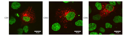
ExoFLARE constructs were transiently transfected into DU145N cells (human prostate cancer cell line). ExoFLARE cell permeable dye was added to the media and cells were imaged using a confocal fluorescence microscope. Red: ExoFLARE-tagged protein (corresponding to exosomes). Green: Hoechst stain (nuclei).
Material available for download
ExoFLARE user guide
