


FLISA, Flow Cytometry, IF Microscopy, Western Blot
Goat
Mus musculus (Mouse)
IgG
Polyclonal
Mouse IgG whole molecule
Goat Anti-Mouse IgG Secondary Antibody fluorescein Conjugated, Goat Anti-Mouse IgG Antibody FITC Conjugated, GAM-FITC, Anti-mouse IgG secondary antibody, anti-mouse IgG Fluorescein conjugated secondary antibody, Gt Anti-Ms IgG FITC Conjugated Antibody
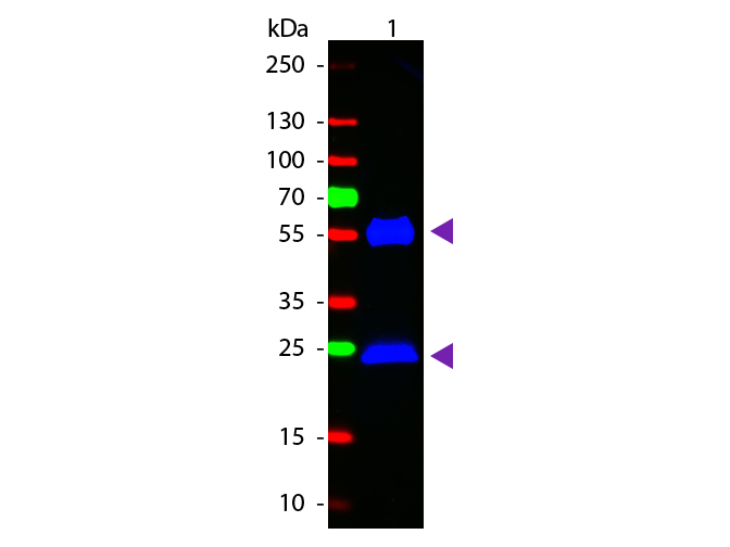
Anti-Mouse IgG [H&L] Fluorescein Antibody generated in goat detects reactivity to Mouse IgG. Secreted as part of the adaptive immune response by plasma B cells, immunoglobulin G constitutes 75% of serum immunoglobulins. Immunoglobulin G binds to viruses, bacteria, as well as fungi and facilitates their destruction or neutralization via agglutination (and thereby immobilizing them) , activation of the compliment cascade, and opsinization for phagocytosis. The whole IgG molecule possesses both the F (c) region, recognized by high-affinity Fc receptor proteins, as well as the F (ab) region possessing the epitope-recognition site. Both the Heavy and Light chains of the antibody molecule are present. Secondary Antibodies are available in a variety of formats and conjugate types. When choosing a secondary antibody product, consideration must be given to species and immunoglobulin specificity, conjugate type, fragment and chain specificity, level of cross-reactivity, and host-species source and fragment composition. Anti-Mouse IgG FITC Conjugated Antibody has been tested by dot blot and western blot and is designed for immunofluorescence microscopy, flow cytometry, fluorescence based plate assays (FLISA) and fluorescent western blotting. This product is also suitable for multiplex analysis, including multicolor imaging, utilizing various commercial platforms.
Store vial at 4°C prior to restoration. For extended storage aliquot contents and freeze at -20°C
