


Applications:
WB, IP
Antibody Host:
Goat
Target:
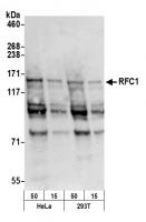
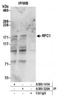
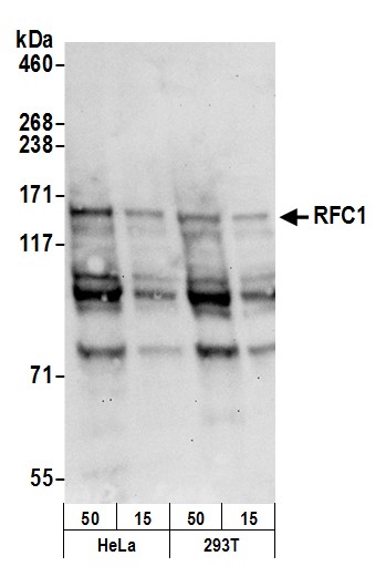
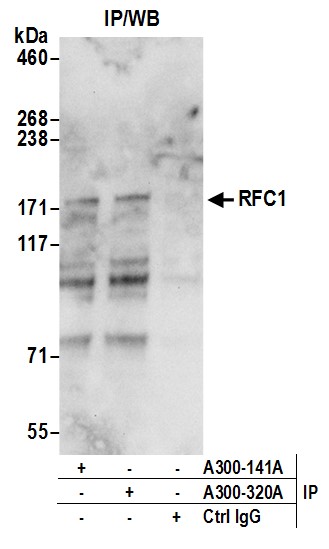
RFC1
Species Reactivity:
Human
Antibody Type:
Polyclonal
Immunogen:
between 1100 and C-term
Alternative Names:
activator 1 140 kDa subunit; replication factor C (activator 1) 1, 145kDa; RFC140; RF-C 140 kDa subunit; RFC; replication factor C1; replication factor C subunit 1; replication factor C large subunit; replication factor C 140 kDa subunit; RECC1; PO-GA; MH
Product Description:
Goat anti-RFC1 Antibody, Affinity Purified - 100 µl (1000 µg/ml)
Storage Temperature:
2 - 8°C

