
Sars-Cov Nonstructural Protein 8 Antibody


IF Microscopy, Immunoprecipitation, Western Blot
Rabbit
SARS Coronavirus nsp8 protein
Antiserum
Polyclonal
This whole rabbit serum was produced by repeated immunizations with a purified His- tagged recombinant protein corresponding to full-length SARS-Coron
rabbit anti-Sars-Cov Nonstructural Protein 8 Antibody, Replicase polyprotein 1a, ORF1a polyprotein, nsp8
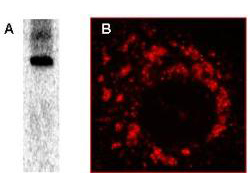
The nonstructural protein 8 (nsp8) is one of the SARS-Coronavirus replicase cleaving products, encoded by ORF1a. Nsp8 is thought to be part of the viral replication complex, which is associated with intracellular membranes. No specific information on the function of nsp8 is available. Anti-SARS-CoV Nonstructural Protein 8 (nsp8) Antibody is useful for researchers interested in viral research. This antibody has been tested for use in immunofluorescence microscopy, immunoelectron microscopy, immunoprecipitation and by western blot. Specific conditions for reactivity should be optimized by the end user. Expect a band of approximately 22 kDa in size corresponding to SARS-CoV nsp8 by western blotting in the appropriate cell lysate or extract. For immunofluorescence microscopy, Vero-E6 cells, grown on glass slides, were infected with SARS-CoV-Fr1 strain for 1 h at 37°C. Infection occurred in PBS/DEAE/2% FCS followed by exchange to EMEM/25mM HEPES/2% FCS. Cells were fixed with PBS/3% PFA. After washing fixed cells, antibody incubation was performed in PBS/5% FCS for 30 min.
Store vial at -20°C prior to opening. Aliquot contents and freeze at -20°C or below for extended sto
