
Rabbit anti-CNOT1 Antibody, Affinity Purified
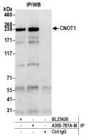
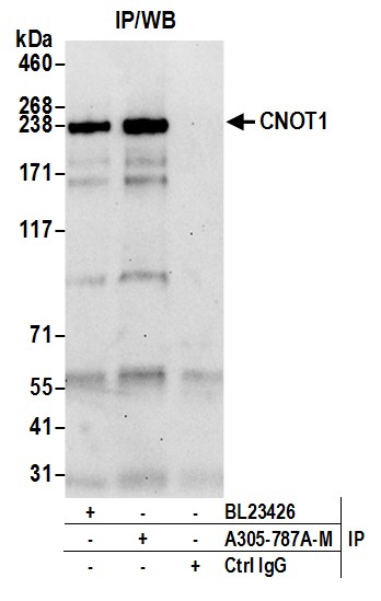
Catalogue number:
A305-787A-M
Supplier:
Size:
100 µl (10 blots) _$$_
Product is available in:


N/A
Shipping is calculated in checkout
This product is no longer available to order.
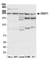
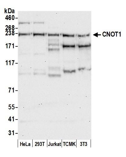
Applications:
WB, IP
Antibody Host:
Rabbit
Species Reactivity:
Human, Mouse
Antibody Isotype:
Whole IgG
Antibody Type:
Polyclonal
Immunogen:
between 1400 and 1450
Alternative Names:
AD-005, adrenal gland protein AD-005, CCR4-associated factor 1, CCR4-NOT transcription complex subunit 1, CDC39, hNOT1, negative regulator of transcription subunit 1 homolog, NOT1, NOT1 (negative regulator of transcription 1, yeast) homolog, NOT1H
Product Description:
Rabbit anti-CNOT1 Antibody, Affinity Purified - 100 µl (10 blots)
Storage Temperature:
2 - 8°C

