


Applications:
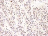
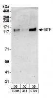
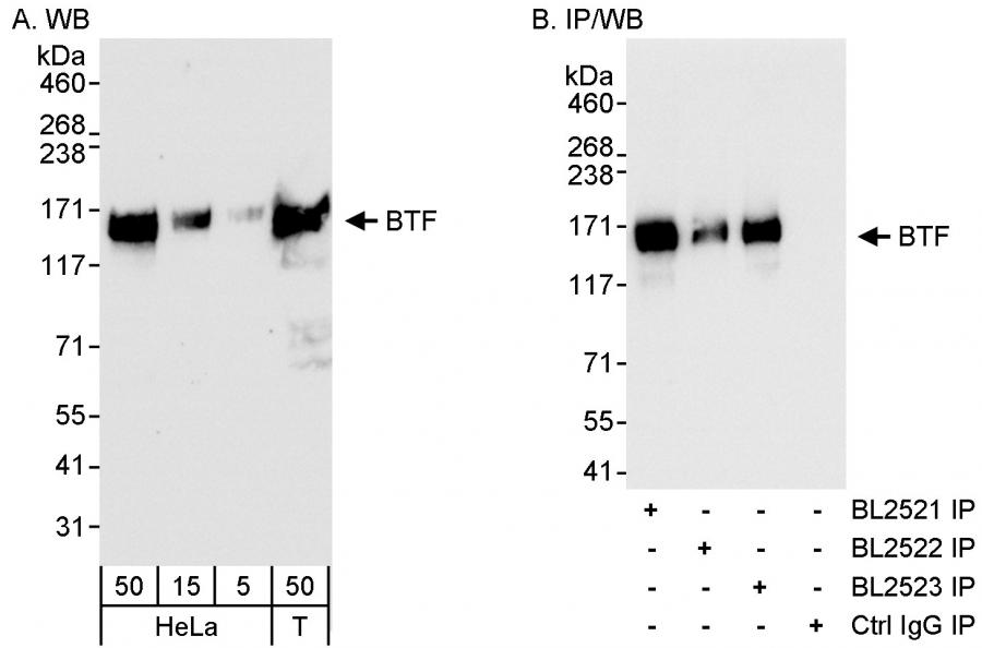
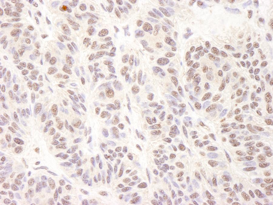
WB, IP, IHC
Antibody Host:
Rabbit
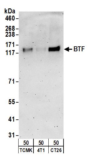
Target:
BTF
Species Reactivity:
Human; Mouse
Antibody Type:
Polyclonal
Immunogen:
between 150 and 200
Alternative Names:
bcl-2-associated transcription factor 1; BTF; bK211L9.1; BCLAF1 and THRAP3 family member 1

Product Description:
Rabbit anti-BTF Antibody, Affinity Purified - 100 µl (200 µg/ml)
Storage Temperature:
2 - 8°C


