
Rabbit anti-TPX2 Antibody, Affinity Purified
Catalogue number:
A300-430A
Supplier:
Size:
100 µl (1000 µg/ml) _$$_
Product is available in:


£436.00
Estimated delivery Fri 26 Apr
Shipping is calculated in checkout
Shipping is calculated in checkout
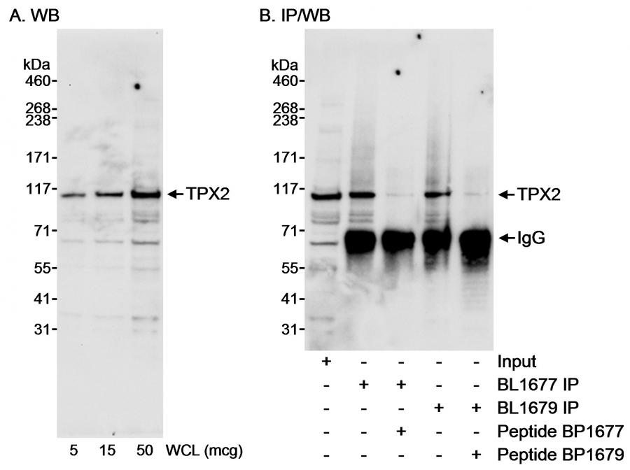
Applications:
WB, IP
Antibody Host:
Rabbit
Target:
TPX2
Species Reactivity:
Human
Antibody Type:
Polyclonal
Immunogen:
between 700 and C-term
Alternative Names:
p100; preferentially expressed in colorectal cancer; protein fls353; REPP86; restricted expression proliferation associated protein 100; Restricted expression proliferation-associated protein 100; targeting protein for Xklp2; TPX2, microtubule-associated
Product Description:
Rabbit anti-TPX2 Antibody, Affinity Purified - 100 µl (1000 µg/ml)
Storage Temperature:
2 - 8°C
