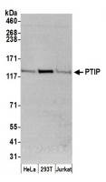
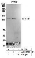
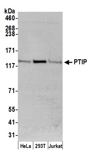
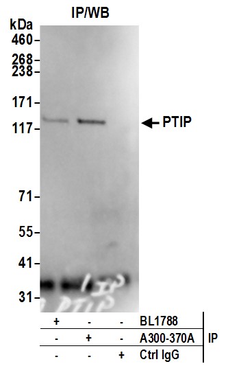
Rabbit anti-PTIP Antibody, Affinity Purified
Antibody was affinity purified using an epitope specific to PTIP immobilized on solid support. The epitope recognized by A300-370A maps to a region between residues 1 and 50 of human PAX transcription activation domain interacting protein 1 using the numbering given in Jowsey, Doherty and Rouse, 2004, J. Biol. Chem. 279(53):55562-55569 (GeneID 22976). Immunoglobulin concentration was determined by extinction coefficient: absorbance at 280 nm of 1.4 equals 1.0 mg of IgG.
- Data sheet/protocol: View or download
- MSDS: View or download
- Applications: WB, IP
Antibodies
CAGF28, CAGF29, PACIP1, PAX interacting (with transcription-activation domain) protein 1, PAX transactivation activation domain-interacting protein, PAX transcription activation domain interacting protein 1 like, PAX-interacting protein 1, PAXIP1L, protei
Human
between 1 and 50
2 - 8°C
Rabbit
Whole IgG
Tris-citrate/phosphate buffer, pH 7 to 8 containing 0.09% Sodium Azide
Polyclonal
12352203

Detection of human PTIP by western blot. Samples: Whole cell lysate (50 µg) from HeLa, HEK293T, and Jurkat cells prepared using NETN lysis buffer. Antibody: Affinity purified rabbit anti-PTIP antibody A300-370A (lot A300-370A-2) used for WB at 0.1 µg/ml. Detection: Chemiluminescence with an exposure time of 30 seconds.

Detection of human PTIP by western blot of immunoprecipitates. Samples: Whole cell lysate (1.0 mg per IP reaction; 20% of IP loaded) from HEK293T cells prepared using NETN lysis buffer. Antibodies: Affinity purified rabbit anti-PTIP antibody A300-370A (lot A300-370A-2) used for IP at 3 µg per reaction. PTIP was also immunoprecipitated by rabbit anti-PTIP antibody BL1788. For blotting immunoprecipitated PTIP, A300-370A was used at 1 µg/ml. Detection: Chemiluminescence with an exposure time of 3 minutes.