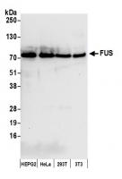
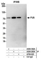
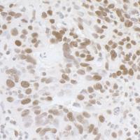
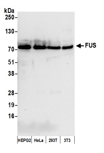
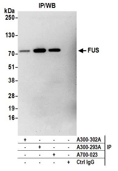
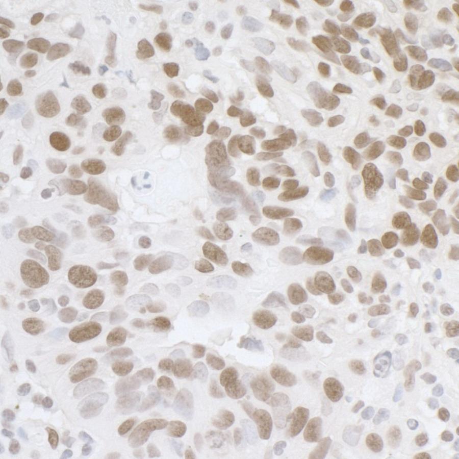
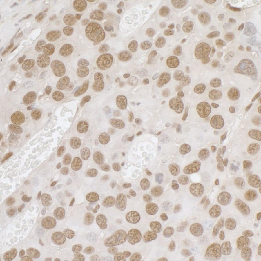
Rabbit anti-FUS Antibody, Affinity Purified - A300-302A - 100 µg - Bethyl Laboratories, Inc. - antibodies


Antibody was affinity purified using an epitope specific to FUS immobilized on solid support. The epitope recognized by A300-302A maps to a region between residues 1 and 50 of human fusion (involved in t(12;16) in malignant liposarcoma) using the numbering given in SwissProt entry P35637 (GeneID 2521). Immunoglobulin concentration was determined by extinction coefficient: absorbance at 280 nm of 1.4 equals 1.0 mg of IgG.
Applications:
WB, IP, IHC

Detection of human and mouse FUS by western blot. Samples: Whole cell lysate (50 µg) from Hep-G2, HeLa, HEK293T, and mouse NIH 3T3 cells prepared using NETN lysis buffer. Antibody: Affinity purified rabbit anti-FUS antibody A300-302A (lot A300-302A-4) used for WB at 0.04 µg/ml. Detection: Chemiluminescence with an exposure time of 10 seconds.

Detection of human FUS by western blot of immunoprecipitates. Samples: Whole cell lysate (1.0 mg per IP reaction; 20% of IP loaded) from HEK293T cells prepared using NETN lysis buffer. Antibodies: Affinity purified rabbit anti-FUS antibody A300-302A (lot A300-293A-4) used for IP at 3 µg per reaction. FUS was also immunoprecipitated by rabbit anti-FUS recombinant monoclonal antibody [BLR023E] (A700-023) and rabbit anti-FUS antibody A300-293A. For blotting immunoprecipitated FUS, A700-023 was used at 1:1000. Chemiluminescence with an exposure time of 3 minutes.

Detection of human FUS by immunohistochemistry. Sample: FFPE section of human ovarian carcinoma. Antibody: Affinity purified rabbit anti-FUS (Cat. No. A300-302A Lot4) used at a dilution of 1:1,000 (1µg/ml). Detection: DAB

Detection of mouse FUS by immunohistochemistry. Sample: FFPE section of mouse renal cells carcinoma. Antibody: Affinity purified rabbit anti-FUS (Cat. No. A300-302A Lot4) used at a dilution of 1:1,000 (1µg/ml). Detection: DAB