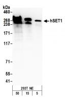
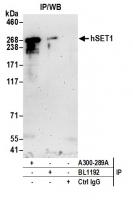
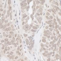
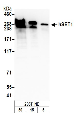
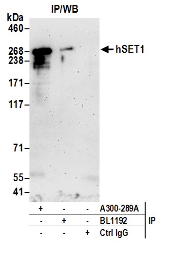
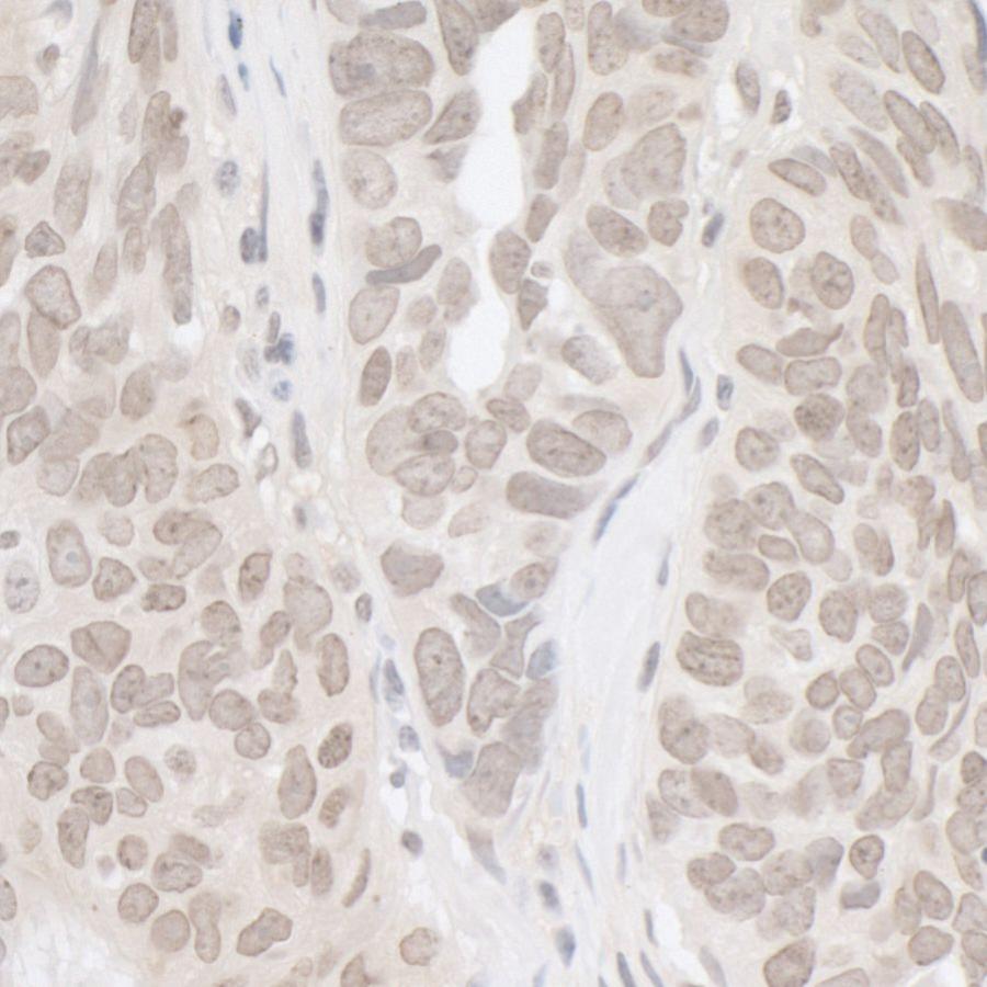
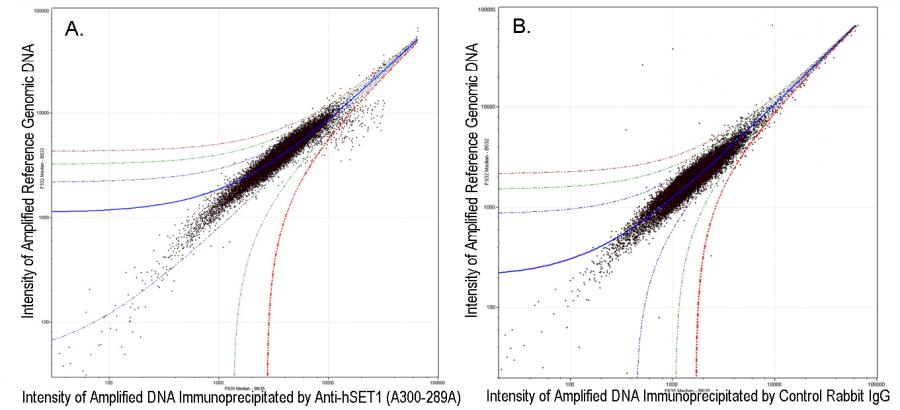
www.bioscience.co.uk
Sales & Support: +44 (0)1223 316 855

*

Rabbit anti-hSET1 Antibody, Affinity Purified

Catalogue number:
Size:
100 µl (1000 µg/ml) _$$_
Product is available in:
  • UK
  • Ireland
  • Europe
  • USA
  • Rest of World
£436.00 Estimated delivery Wed 1 May
Shipping is calculated in checkout
Applications:

WB, IP, IHC

Antibody Host:

Rabbit

Target:

hSET1

Species Reactivity:

Human; Mouse

Antibody Type:

Polyclonal

Immunogen:

between 1200 and 1250

Alternative Names:

Set1A; histone-lysine N-methyltransferase SETD1A; hSET1A; KMT2F; lysine N-methyltransferase 2F; SET domain-containing protein 1A; Set1; set1/Ash2 histone methyltransferase complex subunit SET1

image 0
image 1
image 2
Product Description:

Rabbit anti-hSET1 Antibody, Affinity Purified - 100 µl (1000 µg/ml)

Storage Temperature:

2 - 8°C

Detection of human hSET1 by western blot
Detection of human hSET1 by western blot. Samples: Nuclear extract (50, 15, 5 µg) from HEK293T cells. Antibody: Affinity purified rabbit anti-hSET1 antibody A300-289A (lot A300-289A-4) used for WB at 0.1 µg/ml. Detection: Chemiluminescence with an exposure time of 3 minutes.
Detection of human hSET1 by western blot
Detection of human hSET1 by western blot of immunoprecipitates. Samples: Nuclear Extract (1 mg for IP; 20% of IP loaded) from HEK293T cells. Antibodies: Affinity purified rabbit anti-hSET1 antibody A300-289A (lot A300-289A-4) used for IP at 6 µg per reaction. hSET1 was also immunoprecipitated by rabbit anti-hSET1 antibody BL1192. For blotting immunoprecipitated hSET1, A300-289A was used at 0.4 µg/ml. Detection: Chemiluminescence with an exposure time of 3 minutes.
Detection of human hSet1 by immunohistoc
Detection of human hSet1 by immunohistochemistry. Sample: FFPE section of human ovarian carcinoma. Antibody: Affinity purified rabbit anti-hSet1 Cat. No. A300-289A Lot4 used at a dilution of 1:1,000 (1µg/ml). Detection: DAB
ChIP-chip scatter plot of anti-hSET1 (A3
ChIP-chip scatter plot of anti-hSET1 (A300-289A) enriched DNA binding sites versus input reference DNA. A. 10 µg of A300-289A was used to immunoprecipitate chromatin from K-562 cells according to Ren et al (Genes Dev. 2002 16: 245-256). immunoprecipitatesd DNA and reference DNA were amplified via ligation-mediated PCR and the products labeled with fluorescent dUTPs. The labeled ChIP and reference DNA were pooled, hybridized to a DNA microarray, and analyzed. Data points below the +3 SD curve (red line) represent significantly enriched binding sites. B. As a control, a similar experiment was performed using normal rabbit IgG. Compared to the anti-hSET1 ChIP, normal rabbit IgG showed little enrichment.
Details Cat number & supplier Size Price
SurePAGE™, Bis-Tris, 10x8, 4-12%, 12 wells M00653 · GenScript Biotech
M00653
GenScript Biotech
10 pk/box £56.00
10 pk/box
view
SurePAGE™, Bis-Tris, 10x8, 4-20%, 12 wells M00656 · GenScript Biotech
M00656
GenScript Biotech
10 pk/box £56.00
10 pk/box
view
SurePAGE™, Bis-Tris, 10x8, 10%, 12 wells M00665 · GenScript Biotech
M00665
GenScript Biotech
10 pk/box £56.00
10 pk/box
view