


Applications:
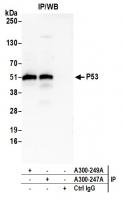
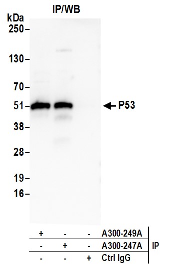
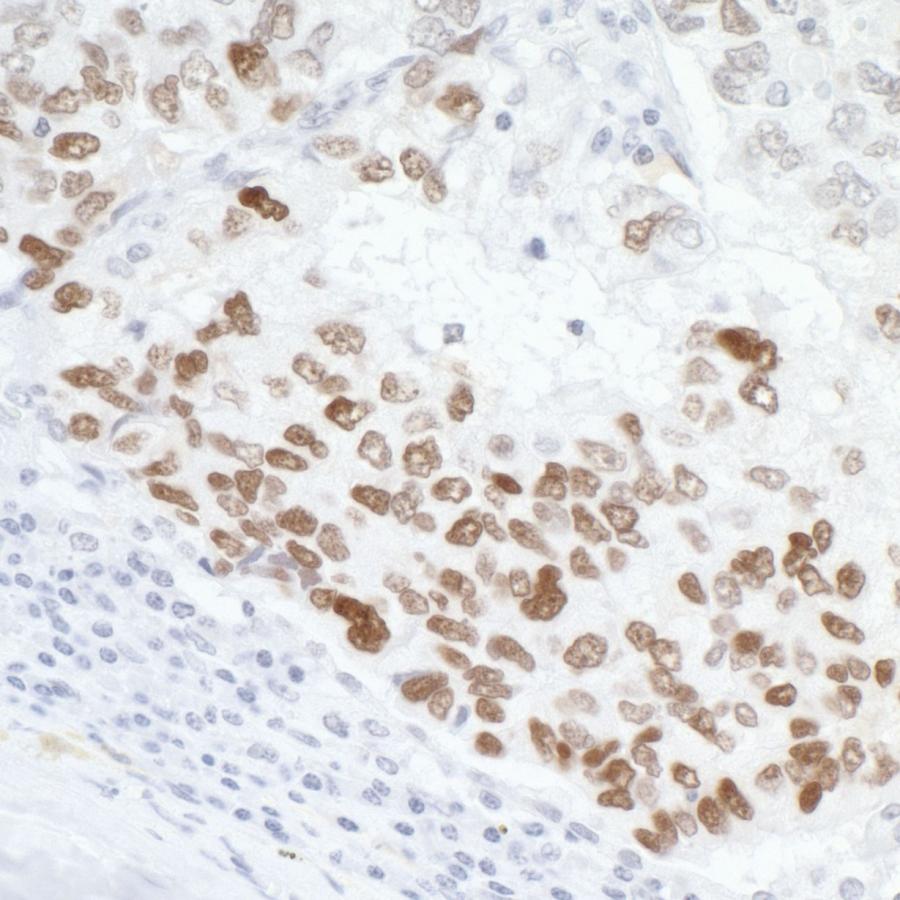
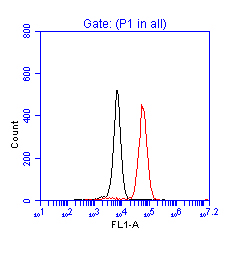
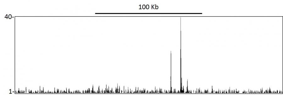
WB, IP, IHC, ChIP-Seq, Flow Cyt
Antibody Host:
Rabbit
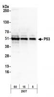
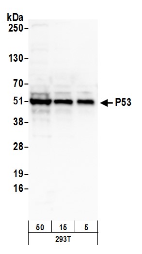
Target:
p53
Species Reactivity:
Human
Antibody Type:
Polyclonal
Immunogen:
between 50 and 100
Alternative Names:
phosphoprotein p53; tumor supressor p53; Tumor suppressor p53; tumor protein 53; TRP53; transformation-related protein 53; P53; mutant tumor protein 53; LFS1; cellular tumor antigen p53; BCC7; antigen NY-CO-13; p53 tumor suppressor

Product Description:
Rabbit anti-p53 Antibody, Affinity Purified - 100 µl (1000 µg/ml)
Storage Temperature:
2 - 8°C


