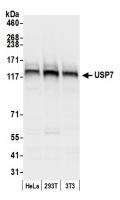
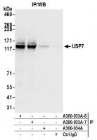
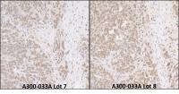
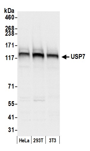
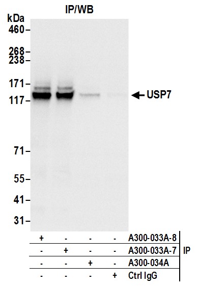
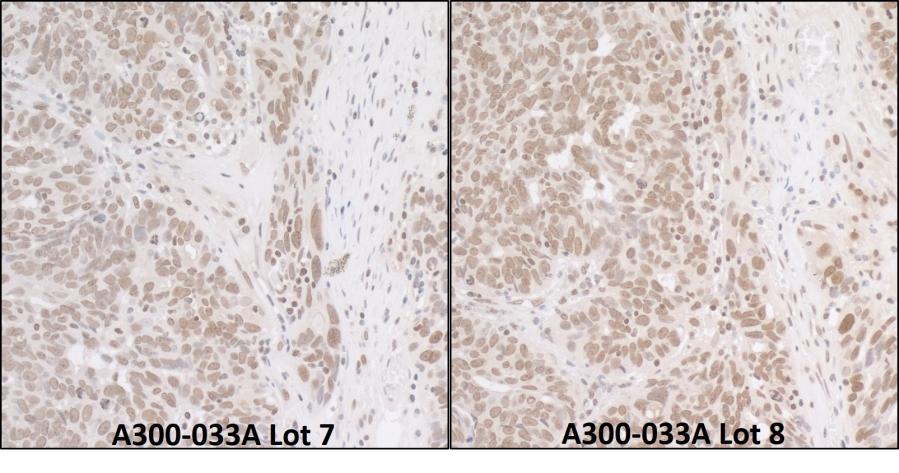
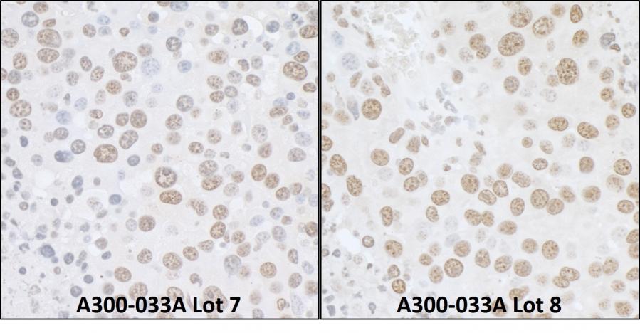
Rabbit anti-USP7 Antibody, Affinity Purified - A300-033A - 100 µg - Bethyl Laboratories, Inc. - antibodies


Antibody was affinity purified using an epitope specific to USP7 immobilized on solid support. The epitope recognized by A300-033A maps to a region between residues 1 and 50 of human Ubiquitin Specific Peptidase 7 using the numbering given in entry NP_003461.1 (GeneID 7874).Immunoglobulin concentration was determined by extinction coefficient: absorbance at 280 nm of 1.4 eq5als 1.0 mg of IgG.
Applications:
WB, IP, IHC

Detection of human and mouse USP7 by western blot. Samples: Whole cell lysate (50 µg) from HeLa, HEK293T, and mouse NIH 3T3 cells prepared using NETN lysis buffer. Antibody: Affinity purified rabbit anti-USP7 antibody A300-033A (lot A300-033A-8) used for WB at 0.1 µg/ml. Detection: Chemiluminescence with an exposure time of 1 second.

Detection of human USP7 by western blot of immunoprecipitates. Samples: Whole cell lysate (0.5 or 1.0 mg per IP reaction; 20% of IP loaded) from HeLa cells prepared using NETN lysis buffer. Antibodies: Affinity purified rabbit anti-USP7 antibody A300-033A (lot A300-033A-8) used for IP at 6 µg per reaction. USP7 was also immunoprecipitated by a previous lot of this antibody (lot A300-033A-7) and rabbit anti-USP7 antibody A300-034A For blotting immunoprecipitated USP7, A300-033A was used at 1 µg/ml. Detection: Chemiluminescence with an exposure time of 1 second.

Detection of human USP7 by immunohistochemistry. Samples: FFPE sections of human ovarian carcinoma. Antibodies: Affinity purified rabbit anti-USP7 Cat. No. A300-033A Lot7 (left) and Lot8 (right) used at a dilution of 1:1,000 (1µg/ml). Detection: DAB

Detection of mouse USP7 by immunohistochemistry. Samples: FFPE sections of mouse renal cell carcinoma. Antibodies: Affinity purified rabbit anti-USP7 Cat. No. A300-033A Lot7 (left) and Lot8 (right) used at a dilution of 1:1,000 (1µg/ml). Detection: DAB