www.bioscience.co.uk
Sales & Support: +44 (0)1223 316 855

*

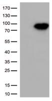
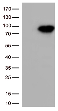
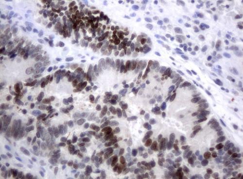
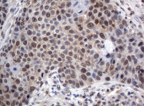
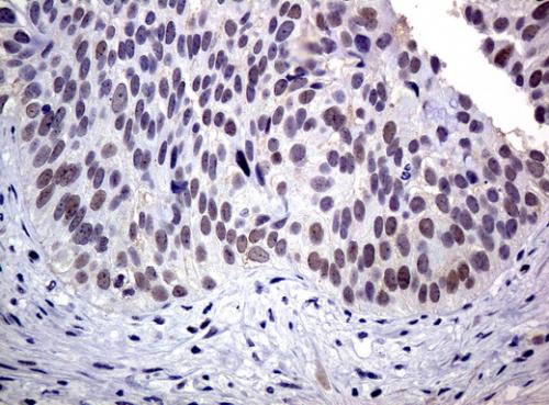
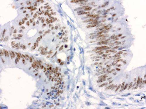
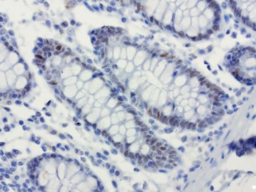
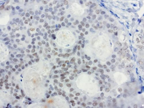
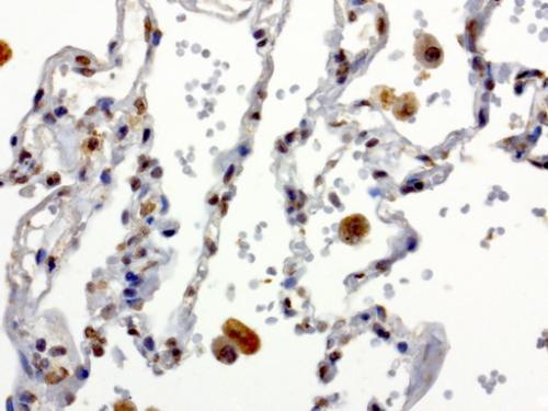
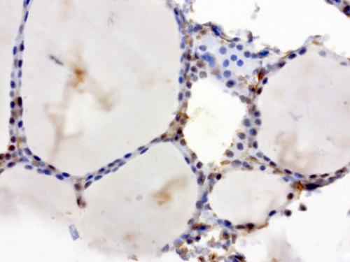
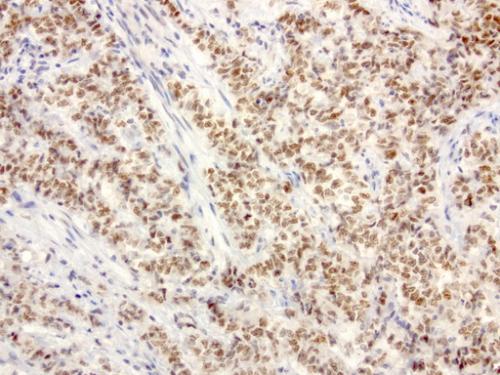
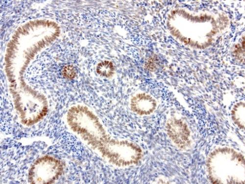
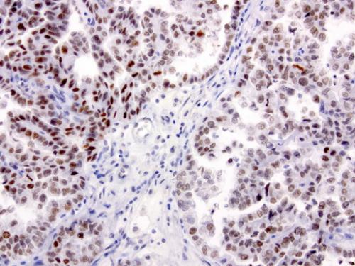
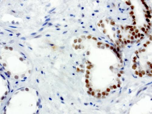
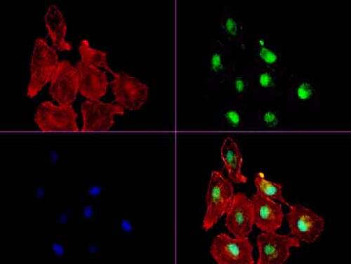
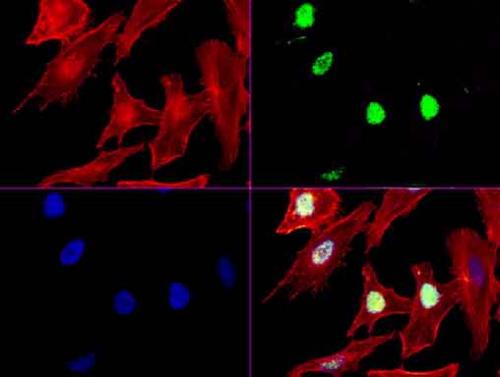
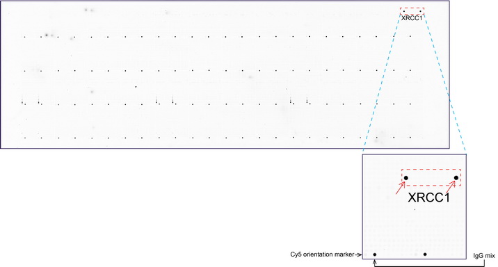
XRCC1 Mouse Monoclonal Antibody [Clone ID: UMAB40]

Catalogue number:
Supplier:
Size:
100 µl _$$_
Product is available in:
  • UK
  • Ireland
  • Europe
  • USA
  • Rest of World
N/A
Shipping is calculated in checkout
This product is no longer available to order.
Applications:

10k-ChIP, IF, IHC, WB

Antibody Host:

Mouse

Species Reactivity:

Human, Monkey, Mouse, Rat

Antibody Isotype:

IgG1

Antibody Type:

Monoclonal

image 0
image 1
image 2
Product Description:

Anti-XRCC1 mouse monoclonal antibody, clone UMAB40

HEK293T cells were transfected with the
HEK293T cells were transfected with the pCMV6-ENTRY control (Left lane) or pCMV6-ENTRY XRCC1 (RC204952, Right lane) cDNA for 48 hrs and lysed. Equivalent amounts of cell lysates (5 ug per lane) were separated by SDS-PAGE and immunoblotted with anti-XRCC1 (1:1000).
Western blot analysis of extracts (35ug)
Western blot analysis of extracts (35ug) from 9 different cell lines by using anti-XRCC1 monoclonal antibody (Clone UMAB40) at 1:500.
Immunohistochemical staining of paraffin
Immunohistochemical staining of paraffin-embedded Adenocarcinoma of colon tissue using anti-XRCC1 mouse monoclonal antibody. (UM500037, dilution 1:50; heat-induced epitope retrieval by 10mM citric buffer, pH6.0, 120°C for 3min)
Immunohistochemical staining of paraffin
Immunohistochemical staining of paraffin-embedded Carcinoma of lung tissue using anti-XRCC1 mouse monoclonal antibody. (UM500037, dilution 1:50; heat-induced epitope retrieval by 10mM citric buffer, pH6.0, 120°C for 3min)
Immunohistochemical staining of paraffin
Immunohistochemical staining of paraffin-embedded Carcinoma of bladder tissue using anti-XRCC1 mouse monoclonal antibody. (UM500037, dilution 1:50; heat-induced epitope retrieval by 10mM citric buffer, pH6.0, 120°C for 3min)
Immunohistochemical staining of paraffin
Immunohistochemical staining of paraffin-embedded human colon carcinoma using anti-XRCC1 clone UMAB40 mouse monoclonal antibody (UM500037) at 1:50 with Polink2 Broad HRP DAB detection kit; heat-induced epitope retrieval with GBI Accel pH8.7 HIER buffer using pressure chamber for 3 minutes at 110C. Nuclear staining is seen in tumor cells.
Immunohistochemical staining of paraffin
Immunohistochemical staining of paraffin-embedded human colon using anti-XRCC1 clone UMAB40 mouse monoclonal antibody (UM500037) at 1:50 with Polink2 Broad HRP DAB detection kit; heat-induced epitope retrieval with GBI Accel pH8.7 HIER buffer using pressure chamber for 3 minutes at 110C. Nuclear staining is seen in epithelial cells.
Immunohistochemical staining of paraffin
Immunohistochemical staining of paraffin-embedded human breast carcinoma using anti-XRCC1 clone UMAB40 mouse monoclonal antibody (UM500037) at 1:50 with Polink2 Broad HRP DAB detection kit; heat-induced epitope retrieval with GBI Accel pH8.7 HIER buffer using pressure chamber for 3 minutes at 110C. Weak nuclear staining is seen in tumor cells.
Immunohistochemical staining of paraffin
Immunohistochemical staining of paraffin-embedded human lung using anti-XRCC1 clone UMAB40 mouse monoclonal antibody (UM500037) at 1:50 with Polink2 Broad HRP DAB detection kit; heat-induced epitope retrieval with GBI TEE pH9.0 HIER buffer using pressure chamber for 3 minutes at 110C. Nuclear staining is seen in epithelial cells.
Immunohistochemical staining of paraffin
Immunohistochemical staining of paraffin-embedded human thyroid using anti-XRCC1 clone UMAB40 mouse monoclonal antibody (UM500037) at 1:50 with Polink2 Broad HRP DAB detection kit; heat-induced epitope retrieval with GBI TEE pH9.0 HIER buffer using pressure chamber for 3 minutes at 110C. Nuclear staining is seen in epithelial cells.
Immunohistochemical staining of paraffin
Immunohistochemical staining of paraffin-embedded human endometrial carcinoma using anti-XRCC1 clone UMAB40 mouse monoclonal antibody (UM500037) at 1:50 with Polink2 Broad HRP DAB detection kit; heat-induced epitope retrieval with GBI TEE pH9.0 HIER buffer using pressure chamber for 3 minutes at 110C. Nuclear staining is seen in tumor cells.
Immunohistochemical staining of paraffin
Immunohistochemical staining of paraffin-embedded human endometrium using anti-XRCC1 clone UMAB40 mouse monoclonal antibody (UM500037) at 1:50 with Polink2 Broad HRP DAB detection kit; heat-induced epitope retrieval with GBI TEE pH9.0 HIER buffer using pressure chamber for 3 minutes at 110C. Nuclear staining is seen in epithelial cells.
Immunohistochemical staining of paraffin
Immunohistochemical staining of paraffin-embedded human ovarian carcinoma using anti-XRCC1 clone UMAB40 mouse monoclonal antibody (UM500037) at 1:50 with Polink2 Broad HRP DAB detection kit; heat-induced epitope retrieval with GBI TEE pH9.0 HIER buffer using pressure chamber for 3 minutes at 110C. Nuclear staining is seen in tumor cells.
Immunohistochemical staining of paraffin
Immunohistochemical staining of paraffin-embedded human prostate carcinoma using anti-XRCC1 clone UMAB40 mouse monoclonal antibody (UM500037) at 1:50 with Polink2 Broad HRP DAB detection kit; heat-induced epitope retrieval with GBI TEE pH9.0 HIER buffer using pressure chamber for 3 minutes at 110C. Nuclear staining is seen in tumor cells.
Immunofluorescent staining of A549 cells
Immunofluorescent staining of A549 cells using anti-XRCC1 mouse monoclonal antibody (UM500037, green). Actin filaments were labeled with TRITC-phalloidin (red), and nuclear with DAPI (blue). The three-color overlay image is located at the bottom-right corner.
Immunofluorescent staining of HeLa cells
Immunofluorescent staining of HeLa cells using anti-XRCC1 mouse monoclonal antibody (UM500037, green). Actin filaments were labeled with TRITC-phalloidin (red), and nuclear with DAPI (blue). The three-color overlay image is located at the bottom-right corner.
OriGene overexpression protein microarra
OriGene overexpression protein microarray chip was immunostained with UltraMAB anti-XRCC1 mouse monoclonal antibody (UM500037). The positive reactive proteins are highlighted with two red arrows in the enlarged subarray. All the positive controls spotted in this subarray are also labeled for clarification. These data show that UltraMAB anti-XRCC1 (Clone UMAB37) very specifically recognizes XRCC1 antigen on OriGene protein microarray chip.
Details Cat number & supplier Size Price
Goat anti-Mouse IgG Heavy and Light Chain Antibody HRP Conjugated A90-116P · Bethyl Laboratories, Inc.
A90-116P
Bethyl Laboratories, Inc.
1 mg £114.00
1 mg
view
Goat anti-Mouse IgG Heavy and Light Chain Cross-Adsorbed Antibody HRP Conjugated A90-216P · Bethyl Laboratories, Inc.
A90-216P
Bethyl Laboratories, Inc.
0.5 mg £124.00
0.5 mg
view