www.bioscience.co.uk
Sales & Support: +44 (0)1223 316 855

*

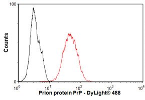
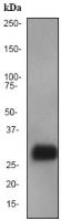
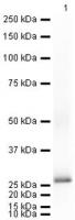
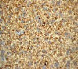
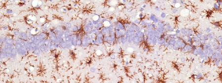
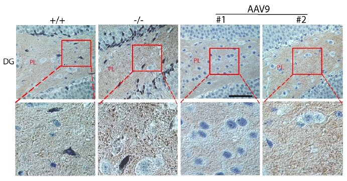
Prion protein PrP (PRNP) Rabbit Monoclonal Antibody [Clone ID: EP1802Y]

Catalogue number:
Supplier:
Size:
100 µl _$$_
Product is available in:
  • UK
  • Ireland
  • Europe
  • USA
  • Rest of World
N/A
Shipping is calculated in checkout
This product is no longer available to order.
Applications:

FC, IHC, WB

Antibody Host:

Rabbit

Species Reactivity:

Human, Mouse, Rat

Antibody Isotype:

IgG

Antibody Type:

Monoclonal

image 0
image 1
image 2
Product Description:

Rabbit Monoclonal Antibody against PRNP (Clone EP1802Y)

Western blot - Prion protein PrP antibod
Western blot - Prion protein PrP antibody [EP1802Y]; Anti-Prion protein PrP antibody [EP1802Y] at 1/10000 dilution + fetal brain lysate at 10 ug.Secondary.HRP-labelled goat anti-rabbit at 1/2000 dilution.Predicted band size : 28 kDa.
Western blot ; Anti-Prion protein PrP an
Western blot ; Anti-Prion protein PrP antibody [EP1802Y] at 1/5000 dilution + Mouse Prion protein PrP full length protein at 0.01 ug.Secondary.Goat polyclonal Secondary Antibody to Rabbit IgG - H&L (HRP), pre-adsorbed at 1/5000 dilution.developed using the ECL technique.Performed under reducing conditions.Exposure time : 10 seconds
Immunohistochemistry (Paraffin-embedded
Immunohistochemistry (Paraffin-embedded sections) - Prion protein PrP antibody [EP1802Y]; Immunohistochemical analysis of brain glioma using TA300958 at a dilution of 1/100-1/250.
Immunohistochemistry (Formalin/PFA-fixed
Immunohistochemistry (Formalin/PFA-fixed paraffin-embedded sections) - Anti-Prion protein PrP antibody [EP1802Y]; Immunohistochemical analysis of Prion-infected mouse brain tissue, staining Prion protein PrP with TA300958. Antigen retrieval was performed by heat mediation in a citrate buffer (pH 6) before incubating with primary antibody (1/7000) overnight at 4°C. Staining was detected using DAB.
Figure from citation: Immunohistochemist
Figure from citation: Immunohistochemistry of PRNP protein level by using anti-PRNP antibody in mouse brain tissue sections. Prnp-positive signals were stained brown. +/+: wt mice; -/-: non-treated MPS IIIB mice; AAV9: rAAV9-treated MPS IIIB mice; #1: mouse with low Prnp IHC intensity; #2: mouse with high Prnp IHC intensity; DG: dentate gyrus of hippocampus. PL: polymoph layer of DG. Scale bar: 50 um. View Citation
Flow Cytometry-Anti-Prion protein PrP an
Flow Cytometry-Anti-Prion protein PrP antibody (TA300958); Overlay histogram showing SH-SY5Y cells stained with TA300958 (red line). The secondary antibody used was DyLight 488 goat anti-rabbit IgG (H+L) at 1:500. Isotype control antibody (black line) was rabbit IgG (monoclonal) (1ug/1x10^6 cells) used under the same conditions. This antibody gave a positive signal in SH-SY5Y cells under the same conditions.
Details Cat number & supplier Size Price
Goat anti-Rabbit IgG Heavy and Light Chain Antibody HRP Conjugated A120-101P · Bethyl Laboratories, Inc.
A120-101P
Bethyl Laboratories, Inc.
1 mg £114.00
1 mg
view
Goat anti-Rabbit IgG Heavy and Light Chain Highly Cross-Adsorbed Antibody HRP Conjugated A120-501P · Bethyl Laboratories, Inc.
A120-501P
Bethyl Laboratories, Inc.
1 mg £194.00
1 mg
view