www.bioscience.co.uk
Sales & Support: +44 (0)1223 316 855

*

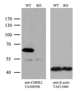
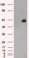
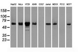
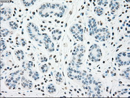
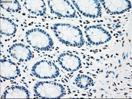
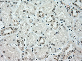
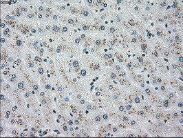
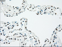
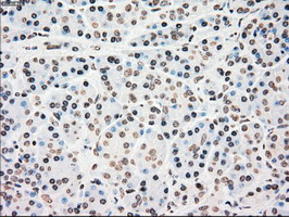
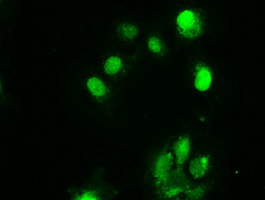
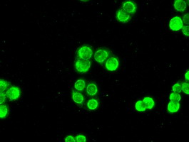
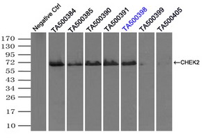
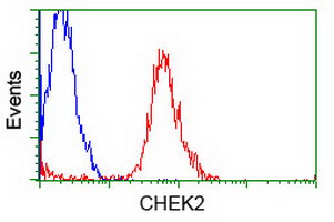
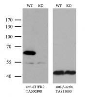
Chk2 (CHEK2) Mouse Monoclonal Antibody [Clone ID: OTI5C4]

Catalogue number:
Supplier:
Size:
100 µl _$$_
Product is available in:
  • UK
  • Ireland
  • Europe
  • USA
  • Rest of World
£533.00 Shipping is calculated in checkout
Applications:

FC, IF, IHC, IP, WB

Antibody Host:

Mouse

Species Reactivity:

Human; Dog; Mouse; Rat

Antibody Isotype:

IgG1

Antibody Type:

Monoclonal

Immunogen:

Full length human recombinant protein of human CHEK2 (NP_009125) produced in HEK293T cell.

Alternative Names:

CDS1; CHK2; hCds1; HuCds1; LFS2; PP1425; RAD53

image 0
image 1
image 2
Product Description:

CHEK2 (CHK2) mouse monoclonal antibody, clone OTI5C4 (formerly 5C4)

Storage Temperature:

Store at -20°C as received.

Equivalent amounts of cell lysates (10 u
Equivalent amounts of cell lysates (10 ug per lane) of wild-type HeLa cells (WT, Cat# LC810HELA) and CHEK2-Knockout hela cells (KO, Cat# LC810324) were separated by SDS-PAGE and immunoblotted with anti-CHEK2 monoclonal antibody TA500398. Then the blotted membrane was stripped and reprobed with anti-β-actin (TA811000) as a loading control (1:500).
HEK293T cells were transfected with the
HEK293T cells were transfected with the pCMV6-ENTRY control (Left lane) or pCMV6-ENTRY CHEK2 (RC201278, Right lane) cDNA for 48 hrs and lysed. Equivalent amounts of cell lysates (5 ug per lane) were separated by SDS-PAGE and immunoblotted with anti-CHEK2. Positive lysates LY416128 (100ug) and LC416128 (20ug) can be purchased separately from OriGene.
Western blot analysis of extracts (35ug)
Western blot analysis of extracts (35ug) from 9 different cell lines by usin g anti-CHEK2 monoclonal antibody (HepG2: human; HeLa: human; SVT2: mouse; A549: human; COS7: monkey; Jurkat: human; MDCK: canine; PC12: rat; MCF7: human).
Immunohistochemical staining of paraffin
Immunohistochemical staining of paraffin-embedded Human breast tissue within the normal limits using anti-CHEK2 mouse monoclonal antibody. (Heat-induced epitope retrieval by 10mM citric buffer, pH6.0, 100°C for 10min, TA500398)
Immunohistochemical staining of paraffin
Immunohistochemical staining of paraffin-embedded Human colon tissue within the normal limits using anti-CHEK2 mouse monoclonal antibody. (Heat-induced epitope retrieval by 10mM citric buffer, pH6.0, 100°C for 10min, TA500398)
Immunohistochemical staining of paraffin
Immunohistochemical staining of paraffin-embedded Human Kidney tissue within the normal limits using anti-CHEK2 mouse monoclonal antibody. (Heat-induced epitope retrieval by 10mM citric buffer, pH6.0, 100°C for 10min, TA500398)
Immunohistochemical staining of paraffin
Immunohistochemical staining of paraffin-embedded Human liver tissue within the normal limits using anti-CHEK2 mouse monoclonal antibody. (Heat-induced epitope retrieval by 10mM citric buffer, pH6.0, 100°C for 10min, TA500398)
Immunohistochemical staining of paraffin
Immunohistochemical staining of paraffin-embedded Human lung tissue within the normal limits using anti-CHEK2 mouse monoclonal antibody. (Heat-induced epitope retrieval by 10mM citric buffer, pH6.0, 100°C for 10min, TA500398)
Immunohistochemical staining of paraffin
Immunohistochemical staining of paraffin-embedded Human pancreas tissue within the normal limits using anti-CHEK2 mouse monoclonal antibody. (Heat-induced epitope retrieval by 10mM citric buffer, pH6.0, 100°C for 10min, TA500398)
Anti-CHEK2 mouse monoclonal antibody (TA
Anti-CHEK2 mouse monoclonal antibody (TA500398) immunofluorescent staining of COS7 cells transiently transfected by pCMV6-ENTRY CHEK2 (RC201278).
Immunofluorescent staining of HT29 cells
Immunofluorescent staining of HT29 cells using anti-CHEK2 mouse monoclonal antibody (TA500398).
Immunoprecipitation (IP) of CHEK2 by usi
Immunoprecipitation (IP) of CHEK2 by using TrueMab monoclonal anti-CHEK2 antibodies (Negative control: IP without adding anti-CHEK2 antibody.). For each experiment, 500ul of DDK tagged CHEK2 overexpression lysates (at 1:5 dilution with HEK293T lysate), 2ug of anti-CHEK2 antibody and 20ul (0.1mg) of goat anti-mouse conjugated magnetic beads were mixed and incubated overnight. After extensive wash to remove any non-specific binding, the immuno-precipitated products were analyzed with rabbit anti-DDK polyclonal antibody.
Flow cytometric Analysis of Jurkat cells
Flow cytometric Analysis of Jurkat cells, using anti-CHEK2 antibody (TA500398), (Red), compared to a nonspecific negative control antibody, (Blue).
Details Cat number & supplier Size Price
Goat anti-Mouse IgG Heavy and Light Chain Antibody HRP Conjugated A90-116P · Bethyl Laboratories, Inc.
A90-116P
Bethyl Laboratories, Inc.
1 mg £114.00
1 mg
view
Goat anti-Mouse IgG Heavy and Light Chain Cross-Adsorbed Antibody HRP Conjugated A90-216P · Bethyl Laboratories, Inc.
A90-216P
Bethyl Laboratories, Inc.
0.5 mg £124.00
0.5 mg
view